Analysis of contaminations on a plastic part using the FT-IR microscope LUMOS
Contaminations of polymer products are often microscopically small and not easy to analyze. Without knowing the chemical composition of the defect, the determination of its origin is often difficult. FT-IR microscopy is an attractive tool for the analysis of contaminations, since this technique is capable of identifying not only organic but also inorganic components. With FT-IR microscopy it is possible to obtain an IR-spectrum anywhere on the sample with high lateral resolution and thereby revealing the chemical composition of a certain area of the sample.
So far most of the commercially available FT-IR microscopes are rather bulky and need an additional FT-IR spectrometer. Moreover they are also quite complex to use and require a high level of skill. With the new FT-IR microscope LUMOS Bruker offers a fully automated stand-alone solution that is very easy to use and requires little lab space.
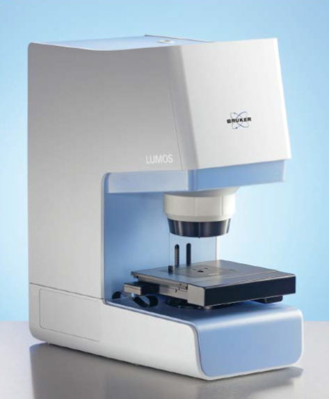 | |
| Figure 1: LUMOS stand-alone FT-IR microscope |
Instrumentation
The very compact FT-IR microscope LUMOS is an all in one solution with an integrated spectrometer, a high degree of motorization and a dedicated user-interface. Its 8x objective provides the measurement modes ATR, transmission and reflection and high quality visual inspection capabilities. The innovative motorized attenuated total reflectance (ATR) crystal allows performing the complete measurement procedure fully automated including background and sample measurements. A high working distance and the unobstructed access to the sample stage facilitate an easy positioning of the sample. The large field of view of 1.5 x 1.2 mm and the high depth of focus make sample inspection very comfortable. In combination with a motorized stage, fully automated mappings can be performed.
The dedicated OPUS Video-wizard is guiding the user through the whole measurement procedure and always provides the appropriate functions for the current measurement step.
Application example: Identification of a contamination on a polycarbonate plastic part
In our example we want to analyze black streaks on a polycarbonate sample shown in figure 2. The question is if the streaks are on the surface or embedded in the PC-matrix and what their chemical nature is. Using the Germanium ATR objective the IR-microscopic analysis can be performed without any sample preparation like the generation of microtome cuts. Moreover Germanium with its high refractive index is an ideal material for the analysis of dark samples with a high surface sensitivity. In addition, the ATR mode provides a lateral resolution limit that is four times better than in transmission or reflection, because the Germanium crystal acts as an immersion lens.
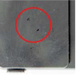 | |
| Figure 2: Macroscopic image of the sample, the black streaks are marked with a circle. |
A total of six spectra were measured on the sample with an acquisition time of 17 seconds per position and a spectral resolution of 4 cm-1. Figure 3 shows the visual image of the contaminated polycarbonate sample with the measurement positions as colored spots. The resulting spectra are shown in figure 4. Those spectra measured on the black contaminations clearly indicate a spectral difference from the spectra taken on the clean polycarbonate.
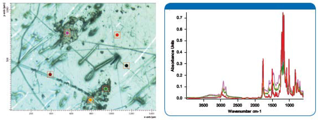 | |
Figure 3: Visual microscopic image of the polycarbonate sample with black streaks. (LUMOS, 8x) |
Since the spectrum of the black spots still contained strong bands of the polycarbonate matrix a difference spectrum was created by subtracting the polycarbonate spectrum (upper spectrum, figure 5) from the spectrum measured on the black spot (middle spectrum, figure 5). The lower spectrum in figure 5 shows the result. A library search revealed that the contaminations originate from a black ink marker (marker ink black 6558). With this information it is now feasible to track down the source of the black streaks.
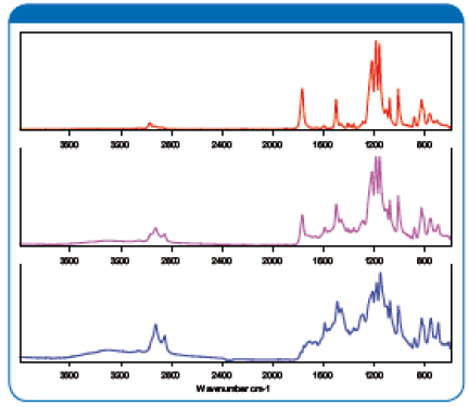 | |
| Figure 5: ATR spectra of the clean polycarbonate matrix (top) and the black spot (middle). Result of the subtraction of the above spectra is shown on the bottom. The library search of the difference spectrum reveals that the contamination originates from a black ink |
Summary
The new LUMOS FT-IR microscope is a compact and fully motorized stand-alone FT-IR microscope with a low cost of ownership. Due to the easy to use OPUS-Video wizard both measurement and analysis can be performed even by spectroscopically inexperienced users. The built in fully motorized ATR objective is an ideal tool for the analysis of surface defects with a high lateral resolution and an outstanding surface sensitivity. In combination with the built in library search function it is possible to quickly identify even unknown samples. Its comfortable use and high performance make the LUMOS an ideal instrument for QC laboratories that have to analyze defects and contaminations in various products on demand. Typical analytical questions like inclusions in polymers and rubbers, impurities on electronic devices and mechanical parts but also in pharmaceutical products can be solved quickly without method specific skills.
- autor:
- O. K. SERVIS BioPro, s. r. o.


